-
作为21世纪三大科学之一的纳米科学,从20世纪80年代以来逐渐成为科学研究的热点. MNPs具有独特的物理化学特性,近年来在多个领域广泛应用,据估计MNPs年产量接近100万吨[1]. 随着MNPs的大量涌现和广泛应用,关于MNPs的环境行为及其生态毒性引起了公众的高度关注. 2003年Science[2]和Nature[3]杂志相继发表文章探讨纳米颗粒存在的安全问题以及对环境和人类健康的影响,从此,MNPs对环境及生物的毒性效应研究逐渐成为国内外的研究热点.
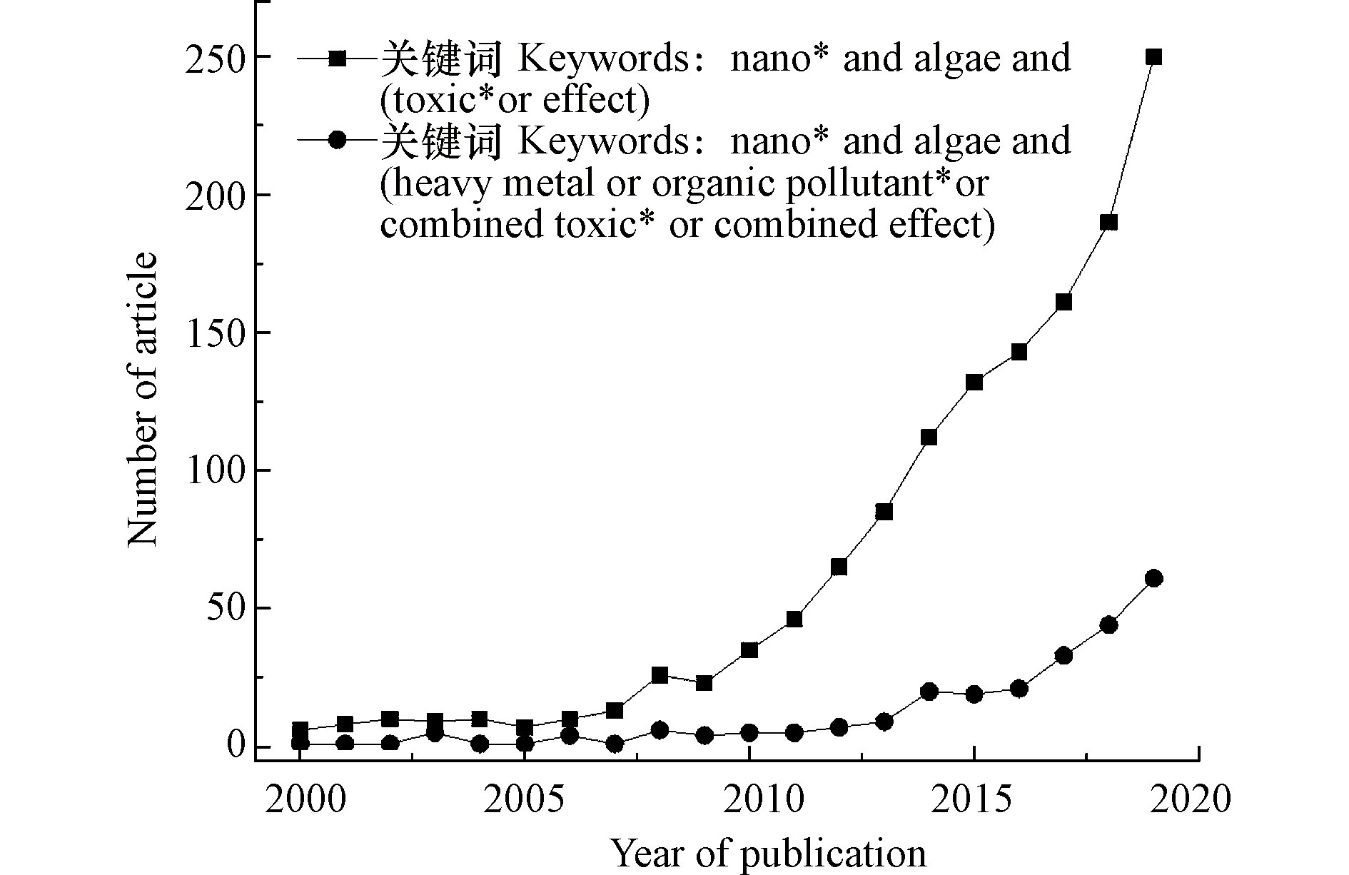
随着MNPs生产和使用的增加,其在环境中的释放量也越来越多,尤其对于水环境,水环境是MNPs的主要汇集地. 在水环境中MNPs会经历各种物理化学反应,且会受到各种因素的影响(如MNPs的物化特性和环境条件).MNPs暴露于水环境中,势必会与水生生物接触,进而被水生生物吸收,并对其产生系列影响. 藻类是水环境中的主要生产者,由于其在MNPs存在时的EC50(在规定暴露时间内产生50%生长抑制率的浓度)值低,即对MNPs敏感性高,而被作为研究MNPs生态影响的典型模型生物之一[4-5]. MNPs被藻类吸收后会随着食物链传递,最终暴露于人类,因此研究MNPs对藻类的毒性效应及效应机制具有重要意义. 近年来MNPs对藻类的毒性研究得到了迅速发展,特别是从2010年以来,相关研究的发文量直线提升,表明MNPs安全性问题特别是藻毒性受到公众的普遍关注(图1).
从图1可以看出,目前已广泛研究单一MNPs对藻类的毒性效应,而尚未充分开展MNPs与环境中共存污染物共同暴露对藻类的复合效应相关研究. 真实环境中,生物往往同时暴露于多种污染物,因此对不同污染物质及多种MNPs共同暴露进行风险评估至关重要[6]. 因此,本文重点介绍了MNPs与环境中共存污染物对藻类的复合效应. 随着组学技术的发展,MNPs对藻类毒性效应的机理研究进入了一个新阶段,蛋白质组学[7]、基因组学[8]、代谢组学[9]、转录组学[10]等组学技术是进一步了解MNPs毒理学的关键,但是目前关于MNPs对藻类的毒性机理的认识并不全面. 此外,对MNPs在环境中的长期动态变化的认知也不够深入. 因此,应该发挥多组学的优势,与传统的生物终端指标结合,探索MNPs的毒性机理,以进一步认识MNPs的安全性问题. 本文系统地综述了MNPs的环境行为、MNPs毒性的影响因素、不同MNPs对藻类的毒性效应和毒性机制、MNPs与环境中共存污染物的联合效应,并对目前相关研究中存在的问题进行了总结及就今后的研究方向进行了展望,希望能有助于理解MNPs在水环境中的风险,促进纳米技术的可持续发展.
人工纳米颗粒对微型藻类的毒性效应及其机制
Toxic effect and mechanism of manufactured nanoparticles on microalgae
-
摘要:
随着纳米技术的飞速发展和人工纳米颗粒(manufactured nanoparticles, MNPs)的广泛应用, 释放到环境中的MNPs不断增多, MNPs的环境行为及生态效应一直是国内外研究的热点. 由于水生生态系统的复杂性, 目前MNPs对藻类的毒性机制仍不明确. 本文以藻类为模型, 综述了MNPs在水环境中的行为, 重点探讨了MNPs的理化性质和环境因子对MNPs生物毒性的影响和调控机制. 系统总结了碳基MNPs、金属基MNPs、量子点及有机聚合物对藻类的致毒效应. 基于复合污染的真实环境, 本文还重点总结了MNPs与环境中共存污染物的复合效应, 并从生物终端指标和组学指标两个方面揭示了MNPs的潜在毒性机制. 最后, 分析了目前纳米毒理学研究中存在的问题, 并对今后的研究方向进行了展望, 以期更好地认识MNPs的环境和生态安全性问题, 以便更好地促进MNPs的可持续发展.
Abstract:With the rapid development of nanotechnology and the wide application of manufactured nanoparticles (MNPs), the large amount of MNPs were released into the environment. The environmental behavior and ecological effects of MNPs have been the hotspots. Due to the complexity of aquatic ecosystem, the toxic mechanism of MNPs on algae is still unclear. In this paper, the behavior of MNPs in the aquatic environment was reviewed. Meanwhile, we emphatically discussed the effect of physical and chemical properties of MNPs and environmental factors on the biological toxicity of MNPs and the regulatory mechanism of algae. The toxic effects of carbon-based MNPs, metal-based MNPs, quantum dots and organic polymer on algae were systematically summarized. Considering the real environment containing various pollutants, this paper also focused on the combined effects of MNPs and coexisting pollutants. The potential toxicity mechanism of MNPs was revealed from two aspects of biological terminal index and omics index. Finally, the existing problems were analyzed in the research of nanotoxicology, and the future research direction was prospected, in order to better understand the environmental and ecological safety of MNPs for promoting the sustainable development of MNPs.
-
Key words:
- manufactured nanoparticles /
- nanotoxicity /
- microalgae /
- oxidative stress /
- combined effects
-
表 1 不同类型MNPs对藻类的毒性效应及其机制
Table 1. Toxic effect and mehanism of different types of MNPs on algae
纳米颗粒
Nanoparticles受试生物
Subject organism毒性效应及机制
Toxic effect and mechanism文献
Reference碳基NPs 绿藻斜角藻 72 h时, 150 mg·L−1和300 mg·L−1的氧化石墨烯(GO)暴露使藻类生长量分别降低了47%和95% [48] 绿藻和蓝藻 蓝藻比绿藻对GO更敏感, 生长抑制更强; 轻度氧化的GO比重度氧化的GO引起的膜损伤严重 [49] 淡水藻类 不同类型石墨烯毒性不同, 还原的氧化石墨烯(rGO)>GO>多层石墨烯(MG),96 h的EC50分别为34.0 mg·L−1、37.3 mg·L−1、62.2 mg·L−1 [50] 小球藻 活性氧产生而导致的细胞膜肿胀, 24 h时促进细胞分裂, 96 h时抑制. 96 h时GO和多壁碳纳米管(SWCNT)对细胞分裂的抑制率分别为0.08%—15%和0.8%—28.3% [51] 斜生栅藻 72 h斜生栅藻对富勒烯(nC60)的亚致死浓度为0.09 mg·L−1,此浓度下, 藻类的光合产物及叶绿素的含量降低 [52] 斜生栅藻 随着nC60浓度增加, 藻类生长速率下降, 浓度>0.18 mg·L−1时, 显著抑制斜生栅藻生长, 72 h的IC50=1.94 mg·L−1 [53] 铜绿假单胞菌和
金黄色葡萄球菌28 d铜绿假单胞菌对nC60的LC50=1336 mg·L−1,而nC60对金黄色葡萄菌群几乎无毒性. 且减少的细菌数量: 富勒烯醇>nC60. 不利影响: 非环氧化nC60−28 d>环氧化nC60−14 d [54] 小球藻 分别将0、5、10 mg·L−1的氧化型多壁碳纳米管(o-MNCNTs)和原始多壁碳纳米管(p-MNCNTs)暴露于小球藻, 发现o-MNCNTs的毒性大于p-MNCNTs, 且急性毒性与CNTs聚集及氧化水平有关 [55] 海藻杜氏藻 0.1—20 mg·L−1时, 单壁碳纳米管的毒性随浓度增加而增强, 生长抑制率高达30%,光和产量下降达18%,谷胱甘肽降低达95% [56] 淡水硅藻 CNT存在下, 24、48、72、96 h的EC50分别为24.64、22.49、28.98、12.15 mg·L−1 [57] 金属基NPs 绿藻 100 mg·L−1的NiO NPs暴露96 h时, 游离的Ni2+为6.42%,EC50=13.7 mg·L−1,藻类细胞暴露于0—100 mg·L−1的NiO NPs, 研究发现浓度越高, 毒性越大, 毒性主要表现为抑制细胞分裂和光合作用的进程、产生ROS、发生氧化应激反应 [5] 绿藻 CeO2 NPs存在下藻类的IC50=7.6—28 mg·L−1,而CeO2 MPs存在下藻类的IC50=59 mg·L−1,表明较小颗粒毒性大. DOM存在下IC50>100 mg·L−1,表明DOM减轻了NPs对藻类的毒性 [23] 短凯伦藻和
中肋骨条藻72 h时对TiO2 NPs的EC50分别为10.69 mg·L−1和7.37 mg·L−1,TiO2 NPs会抑制藻类生长, 发生氧化应激反应, 产生ROS,ROS的产生位置是叶绿体的电子转移链 [58] 小球藻 EC50:ZnO>NiO>CuO>Ti2O3>Fe2O3,ZnO对藻类生长抑制最强, Fe2O3抑制作用最小 [59] 小球藻 分别暴露于0.08、1、10 mg·L−1的Cu2+、CuO NPs和CuO MPs中, 小球藻生长不变、活性氧产生较少、膜损伤明显, 与Cu2+和CuO MPs相比, CuO NPs暴露时, 代谢途径会发生变化, 且具有额外的颗粒特异性毒性 [60] 微藻 20—80 nm 的AgNPs的毒性效应主要由其溶解释放的银离子引起, 而10 nm的AgNPs与Ag+相比由于更容易被生物体利用, 毒性更大 [25] 微囊藻和颤藻 暴露在1—5 mg·L−1的CoNPs下5 d, 微藻的生长速率稳定下降. 5 mg·L−1时, 生物量分别下降78%和88%,且随着浓度越大, 对藻类的毒性越大 [61] 小球藻 带负电的柠檬酸盐包覆的AgNPs(Cit-AgNPs)特异性调节与线粒体功能相关的蛋白质, 破坏相关的代谢途径; 带正点的聚乙烯亚胺包覆的AgNPs(PEI-AgNPs)主要靶向核糖体功能相关蛋白, 中断蛋白合成和遗传信息的传递途径. 另外, AgNPs释放的Ag+影响蛋白质调节和细胞应激的调节 [62] 蓝藻原球藻 AgNPs的浓度>10 μg·L−1时, 藻类数量减少90%以上, 表明此环境中AgNPs的浓度上限为10 μg·L−1,AgNPs对藻类的毒性依赖于细胞密度, 藻毒性主要是由有毒超氧化物的产生和Ag+的浸出引起 [63] 量子点(QDs) 铜绿微囊藻 碳量子点(CQDs)浓度小于50 mg·L−1时, 轻微抑制藻类生长; 浓度大于500 mg·L−1后, 显著抑制藻类生长 [64] 小球藻 无掺杂CQDs, N掺杂CQDs和N、S共掺杂CQDs的96h EC50分别为232.47、185.83、38.56 mg·L−1 [65] 莱茵衣藻 QDs的溶出度随着pH的降低和QDs浓度的增大而增加, 当暴露于QDs时, 生物积累由溶解的Cd2+引起 [66] 聚苯乙烯等其他NPs 微藻 聚苯乙烯(PsNPs)在高于1.8 mg·L−1 时, 使吸收的CO2减少, 促进ROS产生 [67] 绿藻 由于电动电位和细胞膜的变化, PsNPs对绿藻的吸附速率增加 [43] -
[1] KELLER A A, LAZAREVA A. Predicted releases of engineered nanomaterials: From global to regional to local [J]. Environmental Science & Technology Letters, 2013, 1(1): 65-70. [2] SERVICE R F. American chemical society meeting: nanomaterials show signs of toxicity [J]. Science, 2003, 300(5617): 243-243. [3] BRUMFIEL G. Nanotechnology: A little knowledge [J]. Nature, 2003, 424(6946): 246-248. doi: 10.1038/424246a [4] WANG S S, LV J T, MA J Y, et al. Cellular internalization and intracellular biotransformation of silver nanoparticles in Chlamydomonas reinhardtii [J]. Nanotoxicology, 2016, 10(8): 1129-1135. doi: 10.1080/17435390.2016.1179809 [5] OUKARROUM A, ZAIDI W, SAMADANI M, et al. Toxicity of nickel oxide nanoparticles on a freshwater green algal strain of Chlorella vulgaris [J]. BioMed Research International, 2017, 2017: 1-8. [6] ASHAUER R, O'CONNOR I, ESCHER B I. Toxic mixtures in time-the sequence makes the poison [J]. Environmental Science & Technology, 2017, 51(5): 3084-3092. [7] OUYANG S H, ZHOU Q X, ZENG H, et al. Natural nanocolloids mediate the phytotoxicity of graphene oxide [J]. Environmental Science & Technology, 2020, 54(8): 4865-4875. [8] HUANG J, CHENG J P, YI J. Impact of silver nanoparticles on marine diatom Skeletonema costatum [J]. Journal of Applied Toxicology, 2016, 36(10): 1343-1354. doi: 10.1002/jat.3325 [9] LU J, ZHANG S, GAO S H, et al. New insights of the bacterial response to exposure of differently sized silver nanomaterials [J]. Water Research, 2020, 169: 115205. [10] BARROS D, PRADHAN A, PASCOAL C, et al. Transcriptomics reveals the action mechanisms and cellular targets of citrate-coated silver nanoparticles in a ubiquitous aquatic fungus [J]. Environmental Pollution, 2021, 268: 115913. [11] LEAD J R,BATLEY G E, ALVAREZ P J J, et al. Nanomaterials in the environment: Behavior, fate, bioavailability, and effects [J]. Environmental Toxicology and Chemistry, 2012, 31(12): 2893-2096. doi: 10.1002/etc.2027 [12] THOMAS N, KUMAR M, PALMISANO G, et al. Antiscaling 3D printed feed spacers via facile nanoparticle coating for membrane distillation [J]. Water Research, 2021, 189: 116649. [13] MORADI F, SEDAGHAT S, MORADI O, et al. Review on green nano-biosynthesis of silver nanoparticles and their biological activities: With an emphasis on medicinal plants [J]. Inorganic and Nano-Metal Chemistry, 2021, 51(1): 133-142. doi: 10.1080/24701556.2020.1769662 [14] KOOK J K, PHUNG V D, KOH D Y, et al. Facile synthesis of boronic acid-functionalized magnetic nanoparticles for efficient dopamine extraction [J]. Nano Convergence, 2019, 6(1): 30. doi: 10.1186/s40580-019-0200-7 [15] BANIHASHEM S, NIKPOUR NEZHATI M N, PANAHI H A, et al. Synthesis of novel chitosan-g-PNVCL nanofibers coated with gold-gold sulfide nanoparticles for controlled release of cisplatin and treatment of MCF-7 breast cancer [J]. International Journal of Polymeric Materials and Polymeric Biomaterials, 2020, 69(18): 1197-1208. doi: 10.1080/00914037.2019.1683557 [16] VANCE M E, KUIKEN T, VEJERANO E P, et al. Nanotechnology in the real world: Redeveloping the nanomaterial consumer products inventory [J]. Beilstein Journal of Nanotechnology, 2015, 6: 1769-1780. doi: 10.3762/bjnano.6.181 [17] BRATAN S, INSHAKOVA E, INSHAKOV O, et al. World market for nanomaterials: Structure and trends [J]. MATEC Web of Conferences, 2017, 129: 02013. [18] LEAD J R, BATLEY G E, ALVAREZ P J J, et al. Nanomaterials in the environment: Behavior, fate, bioavailability, and effects—An updated review [J]. Environmental Toxicology and Chemistry, 2018, 37(8): 2029-2063. doi: 10.1002/etc.4147 [19] TURAN N B, ERKAN H S, ENGIN G O, et al. Nanoparticles in the aquatic environment: Usage, properties, transformation and toxicity—A review [J]. Process Safety and Environmental Protection, 2019, 130: 238-249. doi: 10.1016/j.psep.2019.08.014 [20] ESPINASSE B P, GEITNER N K, SCHIERZ A, et al. Comparative persistence of engineered nanoparticles in a complex aquatic ecosystem [J]. Environmental Science & Technology, 2018, 52(7): 4072-4078. [21] EVARISTE L, MOTTIER A, LAGIER L, et al. Assessment of graphene oxide ecotoxicity at several trophic levels using aquatic microcosms [J]. Carbon, 2020, 156: 261-271. doi: 10.1016/j.carbon.2019.09.051 [22] GATOO M A, NASEEM S, ARFAT M Y, et al. Physicochemical properties of nanomaterials: implication in associated toxic manifestations [J]. BioMed Research Intnational, 2014, 2014: 498420. [23] ANGEL B M, VALLOTTON P, APTE S C. On the mechanism of nanoparticulate CeO2 toxicity to freshwater algae [J]. Aquatic Toxicology, 2015, 168: 90-97. doi: 10.1016/j.aquatox.2015.09.015 [24] CARLSON C, HUSSAIN S M, SCHRAND A M, et al. Unique cellular interaction of silver nanoparticles: size-dependent generation of reactive oxygen species [J]. The Journal of Physical Chemistry B, 2008, 112(43): 13608-13619. doi: 10.1021/jp712087m [25] IVASK A, KURVET I, KASEMETS K, et al. Size-dependent toxicity of silver nanoparticles to bacteria, yeast, algae, crustaceans and mammalian cells in vitro [J]. PLoS One, 2014, 9(7): e102108. doi: 10.1371/journal.pone.0102108 [26] KALMAN J, PAUL K B, KHAN F R, et al. Characterisation of bioaccumulation dynamics of three differently coated silver nanoparticles and aqueous silver in a simple freshwater food chain [J]. Environmental Chemistry, 2015, 12(6): 662-672. doi: 10.1071/EN15035 [27] FOROOZANDEH P, AZIZ A A. Insight into cellular uptake and intracellular trafficking of nanoparticles [J]. Nanoscale Research Letters, 2018, 13(1): 339. doi: 10.1186/s11671-018-2728-6 [28] ZAMANI H, MORADSHAHI A, JAHROMI H D, et al. Influence of PbS nanoparticle polymer coating on their aggregation behavior and toxicity to the green algae Dunaliella salina [J]. Aquatic Toxicology, 2014, 154: 176-183. doi: 10.1016/j.aquatox.2014.05.012 [29] YUE Y, LI X M, SIGG L , et al. Interaction of silver nanoparticles with algae and fish cells: a side by side comparison [J]. Journal of Nanobiotechnology, 2017, 15(1): 1-11. doi: 10.1186/s12951-017-0254-9 [30] KOSAK NEE ROHDER L A, BRANDT T, SIGG L, et al. Uptake and effects of cerium(Ⅲ) and cerium oxide nanoparticles toChlamydomonas reinhardtii [J]. Aquatic Toxicology, 2018, 197: 41-46. doi: 10.1016/j.aquatox.2018.02.004 [31] SCHIAVO S, OLIVIERO M, MIGLIETTA M, et al. Genotoxic and cytotoxic effects of ZnO nanoparticles for Dunaliella tertiolecta and comparison with SiO2 and TiO2 effects at population growth inhibition levels [J]. Science of the Total Environment, 2016, 550: 619-627. doi: 10.1016/j.scitotenv.2016.01.135 [32] PHAM T L. Effect of silver nanoparticles on tropical freshwater and marine microalgae [J]. Journal of Chemistry, 2019, 2019: 1-7. [33] BUNDSCHUH M, SEITZ F, ROSENFELDT R R, et al. Effects of nanoparticles in fresh waters: Risks, mechanisms and interactions [J]. Freshwater Biology, 2016, 61(12): 2185-2196. doi: 10.1111/fwb.12701 [34] 王震宇, 赵建, 李娜, 等. 人工纳米颗粒对水生生物的毒性效应及其机制研究进展 [J]. 环境科学, 2010, 31(6): 1409-1418. WANG Z Y, ZHAO J, LI N, et al. Review of ecotoxicity and mechanism of engineered nanoparticles to aquatic organisms [J]. Environmental Science, 2010, 31(6): 1409-1418(in Chinese).
[35] VALE G, FRANCO C, DINIZ M S, et al. Bioavailability of cadmium and biochemical responses on the freshwater bivalve Corbicula fluminea—the role of TiO2 nanoparticles [J]. Ecotoxicology and Environmental Safety, 2014, 109: 161-168. doi: 10.1016/j.ecoenv.2014.07.035 [36] SON J, VAVRA J, FORBES V E. Effects of water quality parameters on agglomeration and dissolution of copper oxide nanoparticles (CuO-NPs) using a central composite circumscribed design [J]. Science of the Total Environment, 2015, 521-522: 183-190. doi: 10.1016/j.scitotenv.2015.03.093 [37] ADELEYE A S, KELLER A A. Interactions between algal extracellular polymeric substances and commercial TiO2 nanoparticles in aqueous media [J]. Environmental Science & Technology, 2016, 50(22): 12258-12265. [38] ZHOU C, VITIELLO V, PELLEGRINI D, et al. Toxicological effects of CdSe/ZnS quantum dots on marine planktonic organisms [J]. Ecotoxicology and Environmental Safety, 2016, 123: 26-31. doi: 10.1016/j.ecoenv.2015.09.020 [39] BOOTH A, STORSETH T, ALTIN D, et al. Freshwater dispersion stability of PAA-stabilised cerium oxide nanoparticles and toxicity towards Pseudokirchneriella subcapitata [J]. Science of the Total Environment, 2015, 505: 596-605. doi: 10.1016/j.scitotenv.2014.10.010 [40] KROLL A, BEHRA R, KAEGI R, et al. Extracellular polymeric substances (EPS) of freshwater biofilms stabilize and modify CeO2 and Ag nanoparticles[J]. Plos One, 2014, 9(10): e110709. doi: 10.1371/journal.pone.0110709. [41] MORELLI E, GABELLIERI E, BONOMINI A, et al. TiO2 nanoparticles in seawater: aggregation and interactions with the green alga Dunaliella tertiolecta [J]. Ecotoxicology and Environmental Safety, 2018, 148: 184-193. doi: 10.1016/j.ecoenv.2017.10.024 [42] VERNEUIL L, SILVESTRE J, MOUCHET F, et al. Multi-walled carbon nanotubes, natural organic matter, and the benthic diatom Nitzschia Palea: "A sticky story'' [J]. Nanotoxicology, 2015, 9(2): 219-229. doi: 10.3109/17435390.2014.918202 [43] NOLTE T M, HARTMANN N B, KLEIJN J M, et al. The toxicity of plastic nanoparticles to green algae as influenced by surface modification, medium hardness and cellular adsorption [J]. Aquatic Toxicology, 2017, 183: 11-20. doi: 10.1016/j.aquatox.2016.12.005 [44] GOSWAMI L, KIM K H, DEEP A, et al. Engineered nano particles: Nature, behavior, and effect on the environment [J]. Journal of Environmental Management, 2017, 196: 297-315. [45] 范功端, 陈薇, 郑小梅, 等. 纳米材料对藻细胞毒性效应及致毒机理 [J]. 生态毒理学报, 2018, 13(2): 23-33. doi: 10.7524/AJE.1673-5897.20170420001 FAN G D, CHEN W, ZHENG X M, et al. The cytotoxic effects of nanomaterials on algae and its mechanisms [J]. Asian Journal of Ecotoxicology, 2018, 13(2): 23-33(in Chinese). doi: 10.7524/AJE.1673-5897.20170420001
[46] SENDRA M, YESTE M P, GATICA J M, et al. Direct and indirect effects of silver nanoparticles on freshwater and marine microalgae (Chlamydomonas reinhardtii and Phaeodactylum tricornutum) [J]. Chemosphere, 2017, 179: 279-289. doi: 10.1016/j.chemosphere.2017.03.123 [47] CHEN X J, LU R R, LIU P, et al. Effects of nano-TiO2 on Chlamydomonas reinhardtii cell surface under UV, natural light conditions [J]. Journal of Wuhan University of Technology-Mater. Sci. Ed., 2017, 32(1): 217-222. doi: 10.1007/s11595-017-1583-0 [48] DU S T, ZHANG P, ZHANG R R, et al. Reduced graphene oxide induces cytotoxicity and inhibits photosynthetic performance of the green alga Scenedesmus obliquus [J]. Chemosphere, 2016, 164: 499-507. doi: 10.1016/j.chemosphere.2016.08.138 [49] MALINA T, MARŠáLKOVá E, HOLÁ K, et al. Toxicity of graphene oxide against algae and cyanobacteria: Nanoblade-morphology-induced mechanical injury and self-protection mechanism [J]. Carbon, 2019, 155: 386-396. doi: 10.1016/j.carbon.2019.08.086 [50] ZHAO J, CAO X S, WANG Z Y, et al. Mechanistic understanding toward the toxicity of graphene-family materials to freshwater algae [J]. Water Research, 2017, 111: 18-27. doi: 10.1016/j.watres.2016.12.037 [51] HU X G, OUYANG S H, MU L, et al. Effects of graphene oxide and oxidized carbon nanotubes on the cellular division, microstructure, uptake, oxidative stress, and metabolic profiles [J]. Environmental Science & Technology, 2015, 49(18): 10825-10833. [52] TAO X J, YU Y X, FORTNER J D, et al. Effects of aqueous stable fullerene nanocrystal (nC60) on Scenedesmus obliquus: Evaluation of the sub-lethal photosynthetic responses and inhibition mechanism [J]. Chemosphere, 2015, 122: 162-167. doi: 10.1016/j.chemosphere.2014.11.035 [53] PONTE S, MOORE E A, BORDER C T, et al. Fullerene toxicity in the benthos with implications for freshwater ecosystem services [J]. Science of the Total Environment, 2019, 687: 451-459. doi: 10.1016/j.scitotenv.2019.05.362 [54] INDEGLIA P A, GEORGIEVA A T, KRISHNA V B, et al. Toxicity of functionalized fullerene and fullerene synthesis chemicals [J]. Chemosphere, 2018, 207: 1-9. doi: 10.1016/j.chemosphere.2018.05.023 [55] ZHANG L Q, LEI C, YANG K, et al. Cellular response of Chlorella pyrenoidosa to oxidized multi-walled carbon nanotubes [J]. Environmental Science:Nano, 2018, 5(10): 2415-2425. doi: 10.1039/C8EN00703A [56] THAKKAR M, MITRA S, WEI L P. Effect on growth, photosynthesis, and oxidative stress of single walled carbon nanotubes exposure to marine Alga Dunaliella tertiolecta [J]. Journal of Nanomaterials, 2016: 2016: 1-9. [57] JIA K, SUN C L, WANG Y L, et al. Effect of TiO2 nanoparticles and multiwall carbon nanotubes on the freshwater diatom Nitzschia frustulum: Evaluation of growth, cellular components and morphology [J]. Chemistry and Ecology, 2019, 35(1): 69-85. doi: 10.1080/02757540.2018.1528240 [58] LI F M, LIANG Z, ZHENG X, et al. Toxicity of nano-TiO2 on algae and the site of reactive oxygen species production [J]. Aquatic Toxicology, 2015, 158: 1-13. doi: 10.1016/j.aquatox.2014.10.014 [59] KO K S, KOH D C, KONG I C. Toxicity evaluation of individual and mixtures of nanoparticles based on algal chlorophyll content and cell count [J]. Materials, 2018, 11(1): 121. [60] WANG L, HUANG X L, SUN W L, et al. A global metabolomic insight into the oxidative stress and membrane damage of copper oxide nanoparticles and microparticles on microalga Chlorella vulgaris [J]. Environmental Pollution, 2020, 258: 113647. [61] ANUSHA L. Inhibition effects of cobalt nano particles against fresh water algal blooms caused by Microcystis and Oscillatoria [J]. American Journal of Applied Scientific Research, 2017, 3(4): 26. [62] ZHANG J L, SHEN L, XIANG Q Q, et al. Proteomics reveals surface electrical property-dependent toxic mechanisms of silver nanoparticles in Chlorella vulgaris [J]. Environmental Pollution, 2020, 265: 114743. [63] DEDMAN C J, NEWSON G C, DAVIES G L, et al. Mechanisms of silver nanoparticle toxicity on the marine cyanobacterium Prochlorococcus under environmentally-relevant conditions [J]. Science of the Total Environment, 2020, 747: 141229. [64] YAN Z Y, CHEN J, XIAO A, et al. Effects of representative quantum dots on microorganisms and phytoplankton: A comparative study [J]. RSC Advances, 2015, 5(129): 106406-106412. doi: 10.1039/C5RA23730K [65] XIAO A, WANG C, CHEN J, et al. Carbon and metal quantum dots toxicity on the microalgae Chlorella pyrenoidosa [J]. Ecotoxicology and Environmental Safety, 2016, 133: 211-217. doi: 10.1016/j.ecoenv.2016.07.026 [66] DOMINGOS R F, SIMON D F, HAUSER C, et al. Bioaccumulation and effects of CdTe/CdS quantum dots on Chlamydomonas reinhardtii - nanoparticles or the free ions? [J]. Environmental Science & Technology, 2011, 45(18): 7664-7669. [67] BHATTACHARYA P, LIN S J, TURNER J P, et al. Physical adsorption of charged plastic nanoparticles affects algal photosynthesis [J]. Journal of Physical Chemistry C, 2010, 114(39): 16556-16561. doi: 10.1021/jp1054759 [68] 许志珍, 赵鹏, 张元宝, 等. 人工纳米材料对典型生物的毒性效应研究进展 [J]. 安全与环境学报, 2017, 17(2): 786-792. XU Z Z, ZHAO P, ZHANG Y B, et al. Research progress review in the toxic effects of the engineering nanomaterials on the typical organisms [J]. Journal of Safety and Environment, 2017, 17(2): 786-792(in Chinese).
[69] YANG W W, MIAO A J, YANG L Y. Cd2+ Toxicity to a green alga Chlamydomonas reinhardtii as influenced by its adsorption on TiO2 engineered nanoparticles [J]. PLoS One, 2012, 7(3): e32300. doi: 10.1371/journal.pone.0032300 [70] SU Y, YAN X M, PU Y B, et al. Risks of single-walled carbon nanotubes acting as contaminants-carriers: potential release of phenanthrene in Japanese medaka (Oryzias latipes) [J]. Environmental Science & Technology, 2013, 47(9): 4704-4710. [71] QU R J, WANG X H, WANG Z Y, et al. Metal accumulation and antioxidant defenses in the freshwater fish Carassius auratus in response to single and combined exposure to cadmium and hydroxylated multi-walled carbon nanotubes [J]. Journal of Hazardous Materials, 2014, 275: 89-98. doi: 10.1016/j.jhazmat.2014.04.051 [72] LAMMEL T, BOISSEAUX P, NAVAS J M. Potentiating effect of graphene nanomaterials on aromatic environmental pollutant-induced cytochrome P450 1A expression in the topminnow fish hepatoma cell line PLHC-1 [J]. Environmental Toxicology, 2015, 30(10): 1192-1204. doi: 10.1002/tox.21991 [73] CHEN Q Q, YIN D Q, HU X L, et al. The effect of nC60 on tissue distribution of ibuprofen in Cyprinus carpio [J]. Science of the Total Environment, 2014, 496: 453-460. doi: 10.1016/j.scitotenv.2014.07.074 [74] HARTMANN N B, von der KAMMER F, HOFMANN T, et al. Algal testing of titanium dioxide nanoparticles—testing considerations, inhibitory effects and modification of cadmium bioavailability [J]. Toxicology, 2010, 269(2-3): 190-197. doi: 10.1016/j.tox.2009.08.008 [75] DALAI S, PAKRASHI S, BHUVANESHWARI M, et al. Toxic effect of Cr(Ⅵ) in presence of n-TiO2 and n-Al2O3 particles towards freshwater microalgae [J]. Aquatic Toxicology, 2014, 146: 28-37. doi: 10.1016/j.aquatox.2013.10.029 [76] CHEN J Y, QIAN Y, LI H R, et al. The reduced bioavailability of copper by nano-TiO2 attenuates the toxicity to Microcystis aeruginosa [J]. Environmental Science and Pollution Research, 2015, 22(16): 12407-12414. doi: 10.1007/s11356-015-4492-9 [77] TANG Y L, LI S Y, QIAO J L, et al. Synergistic effects of nano-sized titanium dioxide and zinc on the photosynthetic capacity and survival of Anabaena sp [J]. International Journal of Molecular Sciences, 2013, 14(7): 14395-14407. doi: 10.3390/ijms140714395 [78] TANG Y L, TIAN J L, LI S Y, et al. Combined effects of graphene oxide and Cd on the photosynthetic capacity and survival of Microcystis aeruginosa [J]. Science of the Total Environment, 2015, 532: 154-161. doi: 10.1016/j.scitotenv.2015.05.081 [79] ZHANG L Q, LEI C, CHEN J J, et al. Effect of natural and synthetic surface coatings on the toxicity of multiwalled carbon nanotubes toward green algae [J]. Carbon, 2015, 83: 198-207. doi: 10.1016/j.carbon.2014.11.050 [80] LIU N, WANG Y P, GE F, et al. Antagonistic effect of nano-ZnO and cetyltrimethyl ammonium chloride on the growth of Chlorella vulgaris: Dissolution and accumulation of nano-ZnO [J]. Chemosphere, 2018, 196: 566-574. doi: 10.1016/j.chemosphere.2017.12.184 [81] 陈昊喆. 纳米ZnO/十二烷基苯磺酸钠复合污染体系对小球藻生长的影响[D]. 湘潭: 湘潭大学, 2018. CHEN H Z. The joint effect of binary mixtures of nano-ZnO and sodium dodecyl benzene sulfonate on the growth of Chlorella vulgaris[D]. Xiangtan: Xiangtan University, 2018 (in Chinese).
[82] EVERETT W N, CHERN C, SUN D Z, et al. Phosphate-enhanced cytotoxicity of zinc oxide nanoparticles and agglomerates [J]. Toxicology Letters, 2014, 225(1): 177-184. doi: 10.1016/j.toxlet.2013.12.005 [83] ISWARYA V, JOHNSON J B, PARASHAR A, et al. Modulatory effects of Zn2+ ions on the toxicity of citrate- and PVP-capped gold nanoparticles towards freshwater algae, Scenedesmus obliquus [J]. Environmental Science and Pollution Research, 2017, 24(4): 3790-3801. doi: 10.1007/s11356-016-8131-x [84] LI L, FERNANDEZ-CRUZ M L, CONNOLLY M, et al. The potentiation effect makes the difference: non-toxic concentrations of ZnO nanoparticles enhance Cu nanoparticle toxicity in vitro [J]. Science of the Total Environment, 2015, 505: 253-260. doi: 10.1016/j.scitotenv.2014.10.020 [85] YE N, WANG Z, FANG H, et al. Combined ecotoxicity of binary zinc oxide and copper oxide nanoparticles to Scenedesmus obliquus [J]. Journal of Environmental Science and Health, 2017, 52(6): 555-560. doi: 10.1080/10934529.2017.1284434 [86] YE N, WANG Z, WANG S, et al. Toxicity of mixtures of zinc oxide and graphene oxide nanoparticles to aquatic organisms of different trophic level: Particles outperform dissolved ions [J]. Nanotoxicology, 2018, 12(5): 423-438. doi: 10.1080/17435390.2018.1458342 [87] TONG T Z, WILKE C M, WU J S, et al. Combined Toxicity of Nano-ZnO and Nano-TiO2: from single to multinanomaterial systems [J]. Environmental Science & Technology, 2015, 49(13): 8113-8123. [88] WILKE C M, TONG T Z, GAILLARD J F, et al. Attenuation of microbial stress due to nano-Ag and nano-TiO2 interactions under dark conditions [J]. Environmental Science & Technology, 2016, 50(20): 11302-11310. [89] WILKE C M, WUNDERLICH B, GAILLARD J F, et al. Synergistic bacterial stress results from exposure to nano-Ag and nano-TiO2 mixtures under light in environmental media [J]. Environmental Science & Technology, 2018, 52(5): 3185-3194. [90] ZHANG C, CHEN X H, TAN L J, et al. Combined toxicities of copper nanoparticles with carbon nanotubes on marine microalgae Skeletonema costatum [J]. Environmental Science and Pollution Research, 2018, 25(13): 13127-13133. doi: 10.1007/s11356-018-1580-7 [91] HUYNH K A, MCCAFFERY J M, CHEN K L. Heteroaggregation reduces antimicrobial activity of silver nanoparticles: Evidence for nanoparticle-cell proximity effects [J]. Environmental Science & Technology Letters, 2014, 1(9): 361-366. [92] HUANG B, WEI Z B, YANG L Y, et al. Combined toxicity of silver nanoparticles with hematite or plastic nanoparticles toward two freshwater algae [J]. Environmental Science & Technology, 2019, 53(7): 3871-3879. [93] HUANG B, YAN S, XIAO L, et al. Label-free imaging of nanoparticle uptake competition in single cells by hyperspectral stimulated raman scattering [J]. Small, 2018, 14(10): 1703246. [94] LIU Y H, WANG S, WANG Z, et al. TiO2, SiO2 and ZrO2 nanoparticles synergistically provoke cellular oxidative damage in freshwater microalgae [J]. Nanomaterials (Basel), 2018, 8(2): 95. [95] QUIGG A, CHIN W C, CHEN C S, et al. Direct and indirect toxic effects of engineered nanoparticles on algae: role of natural organic matter [J]. ACS Sustainable Chemistry & Engineering, 2013, 1(7): 686-702. [96] CHEN F R, XIAO Z G, YUE L, et al. Algae response to engineered nanoparticles: current understanding, mechanisms and implications [J]. Environmental Science-Nano, 2019, 6(4): 1026-1042. doi: 10.1039/C8EN01368C [97] von MOOS N, SLAVEYKOVA V I. Oxidative stress induced by inorganic nanoparticles in bacteria and aquatic microalgae—state of the art and knowledge gaps [J]. Nanotoxicology, 2014, 8(6): 605-630. doi: 10.3109/17435390.2013.809810 [98] DUAN G X, ZHANG Y Z, LUAN B Q, et al. Graphene-induced pore formation on cell membranes [J]. Scientific Reports, 2017, 7: 42767. [99] ZHANG J L, ZHOU Z P, PEI Y, et al. Metabolic profiling of silver nanoparticle toxicity inMicrocystis aeruginosa [J]. Environmental Science:Nano, 2018, 5(11): 2519-2530. [100] GRZELCZAK M P, DANKS S P, KLIPP R C, et al. Ion transport across biological membranes by carborane-capped gold nanoparticles [J]. Acs Nano, 2017, 11(12): 12492-12499. doi: 10.1021/acsnano.7b06600 [101] SENDRA M, BLASCO J, ARAUJO C V M. Is the cell wall of marine phytoplankton a protective barrier or a nanoparticle interaction site? Toxicological responses of Chlorella autotrophica and Dunaliella salina to Ag and CeO2 nanoparticles [J]. Ecological Indicators, 2018, 95: 1053-1067. doi: 10.1016/j.ecolind.2017.08.050 [102] NGUYEN M K, MOON J Y, LEE Y C. Microalgal ecotoxicity of nanoparticles: An updated review [J]. Ecotoxicology and Environmental Safety, 2020, 201: 110781. doi: 10.1016/j.ecoenv.2020.110781 [103] LI X J, SUN H, MAO X M, et al. Enhanced photosynthesis of carotenoids in microalgae driven by light-harvesting gold nanoparticles [J]. ACS Sustainable Chemistry & Engineering, 2020, 8(20): 7600-7608. [104] QIAN H F, ZHU K, LU H P, et al. Contrasting silver nanoparticle toxicity and detoxification strategies in Microcystis aeruginosa and Chlorella vulgaris: New insights from proteomic and physiological analyses [J]. Science of the Total Environment, 2016, 572: 1213-1221. doi: 10.1016/j.scitotenv.2016.08.039 [105] LEI C, ZHANG L Q, YANG K, et al. Toxicity of iron-based nanoparticles to green algae: Effects of particle size, crystal phase, oxidation state and environmental aging [J]. Environmental Pollution, 2016, 218: 505-512. doi: 10.1016/j.envpol.2016.07.030 [106] TAYLOR N S, MERRIFIELD R, WILLIAMS T D, et al. Molecular toxicity of cerium oxide nanoparticles to the freshwater alga Chlamydomonas reinhardtii is associated with supra-environmental exposure concentrations [J]. Nanotoxicology, 2016, 10(1): 32-41. [107] MIDDEPOGU A, HOU J, GAO X, et al. Effect and mechanism of TiO2 nanoparticles on the photosynthesis ofChlorella pyrenoidosa [J]. Ecotoxicology and Environmental Safety, 2018, 161: 497-506. doi: 10.1016/j.ecoenv.2018.06.027 [108] YU Z, ZHANG T, ZHU Y. Whole-genome re-sequencing and transcriptome reveal cadmium tolerance related genes and pathways in Chlamydomonas reinhardtii [J]. Ecotoxicology and Environmental Safety, 2020, 191: 110231. doi: 10.1016/j.ecoenv.2020.110231 [109] TAYLOR C, MATZKE M, KROLL A, et al. Toxic interactions of different silver forms with freshwater green algae and cyanobacteria and their effects on mechanistic endpoints and the production of extracellular polymeric substances [J]. Environmental Science-Nano, 2016, 3(2): 396-408. doi: 10.1039/C5EN00183H [110] von MOOS N, BOWEN P, SLAVEYKOVA V I. Bioavailability of inorganic nanoparticles to planktonic bacteria and aquatic microalgae in freshwater [J]. Environmental Science:Nano, 2014, 1(3): 214-232. doi: 10.1039/c3en00054k [111] HE M L, YAN Y Q, PEI F, et al. Improvement on lipid production by Scenedesmus obliquus triggered by low dose exposure to nanoparticles [J]. Scientific Reports, 2017, 7(1): 15526. doi: 10.1038/s41598-017-15667-0